Nuclear Medicine and PET/CT

Nuclear Medicine and PET/CT

Using small amounts of radioactivity in the form of radiopharmaceuticals, nuclear medicine exams provide valuable information about the function of organs and tissues. PET/CT, a subspecialty of nuclear medicine, provides images that represent the metabolic processes of your tissues. 

Nuclear Medicine

Nuclear medicine uses small amounts of radioactive material, called radiopharmaceuticals, to produce diagnostic images that reveal how organs and tissues are functioning. These radiopharmaceuticals target specific areas of the body. Once administered, a gamma camera detects and translates the radiation to create images. This allows physicians to evaluate organ function, detect abnormalities, and, in some cases, target and destroy diseased cells.

PET/CT

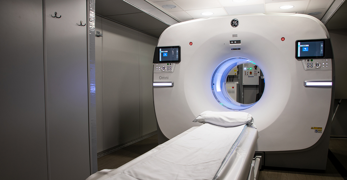
Positron emission tomography (PET) combined with computed tomography (CT) is a specialized form of nuclear medicine imaging. PET/CT scans use radiopharmaceuticals, also known as tracers, to show the metabolic activity of tissues and organs. The tracers emit a type of radiation called positron emission, which is captured by a PET/CT scanner. This technology creates images that reflect biological processes, giving physicians valuable insights into the body’s cellular activity.